Maintaining a working mental representation of this environment to augment one’s capabilities during operative intervention can be particularly cumbersome.You can now actually be able to explore the 3D visualization of the human anatomy with this great Augmented and also a Virtual Reality infused app which is called papar human anatomy.with this wonderful app you can also be able to Disassemble all the major bodily systems, you can do that while you are actually learning about the each individual components functionality and also its. Images and content are created by faculty, staff, and students at An understanding of skull anatomy is important for neurosurgical efficienty. The advent of virtual reality and advances in computer graphics technology has enabled the development of simulated experiences and illustrative representations of intricate anatomical relationships. If you have problems using this site, or have other questions, please feel free to contact us. Previously, all the area between the bones of the rib cage would be filled with noisy data and artifacts which you would need to clean up meticulously.eSkeletons provides an interactive environment in which to examine and learn about skeletal anatomy through our osteology database. What makes this model special is the ultra-high level of detail and the incredible cleanliness of data that the scanner is able to achieve, all thanks to HD Mode. This human skeleton 3D model was captured with an Artec Eva 3D scanner and the newly released AI-powered HD Mode of Artec Studio 15.
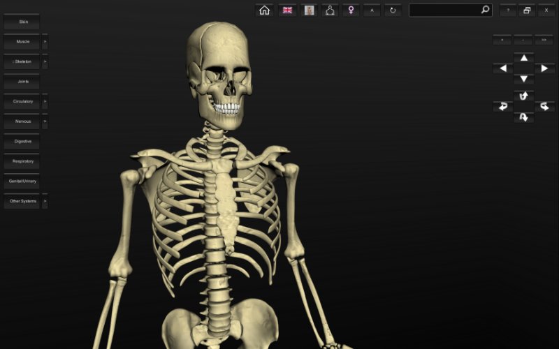
3D Interactive Human Skeleton Full Anatomical Relationships
For educational institutions an excellent opportunity to conduct exciting presentation with excellent imaging. Therefore, there is a need for models that can assist with 3D mental reconstruction of neuroanatomical principles for understanding both normal and pathological cerebral structures.You can also access our Visible Body online databases to view body systems and 3D models of the human body: Visible Body Anatomy & Physiology has the.Complete Human Anatomy Primal 3D Interactive - is not just an atlas or a set of high-quality images - this is 'live' animated projects, with which it is possible to demonstrate disease, trauma, as well as options for their prevention. Based on these resources, surgeons have to reconstruct the anatomy in the three-dimensional (3D) space to appreciate the full anatomical relationships.
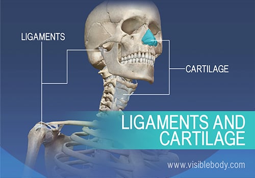
Cranial digital surgical simulation was first initiated in the late 1980’s and early 1990’s. 1,2 This endeavour introduced 3D modelling as a novel means of referencing anatomical data.Digital modelling technology is particularly useful for the field of neurosurgery given the intricate 3D anatomy within the cranial contents and spine. The Visible Human Project was an endeavour by the National Library of Medicine to create a complete 3D representation of a male and female human body for the purpose of education. Did you know The human skeleton consists of both fused and individual bones.Computer graphics and 3D digital designs have an established presence in the neurosurgical literature, particularly within the past decade, to augment education.

The squamous portion is the largest and smoothest. 21,22 The frontal bone is made up of three parts: the squamous, orbital and nasal parts. The frontal bone articulates with the right and left parietal bones, the zygomatic bones, the sphenoid bone, the ethmoid bones, lacrimal bones, maxillary bones, and the nasal bones. We believe these models and accompanying text will provide a useful reference for neurosurgical applications.The frontal bone is a large, unpaired bone that starts out developmentally as two halves that fuse together, along the metopic suture. The virtual human skull model has been divided into 6 different anatomical zones to facilitate illustration of the intricate anatomical relationships.
23Inferior to the glabella lie the nasal notch and spine, which articulate with the nasal bones and the perpendicular plate of the ethmoid. Laterally, the supraorbital margins form the orbital rim and contain the supraorbital notch which transmits the supraorbital vessels and nerves. 21,22 Beneath these are two superciliary arches joined in the middle by the glabella.
23 The orbital portion of the frontal bone contains the frontal sinuses and the frontonasal ducts.The ethmoid bone is an unpaired bone shaped like a cube that articulates with 13 cranial and facial bones. 22,23 The inferior surface of each orbital plate contains a small depression under the zygomatic process called the lacrimal fossa. The orbital portion of the frontal bone is formed by two orbital plates joined by the ethmoidal notch, which is filled by the cribriform plate of the ethmoid. The edges of the sulcus extend inferiorly to form the frontal crest, to which the falx cerebri attaches.
Anteriorly, this articulation forms the foramen cecum. 22 The cribriform plate integrates into the ethmoidal notch of the frontal bone. The ethmoid has three parts: the cribriform plate, the ethmoidal labyrinth, and perpendicular plate.

25 The greater wings make up the anterior portions of both middle fossae and the lesser wings make up the posterior portion of the anterior cranial fossa. 22,24 This bone is the center of attention in endonasal skull base surgery.The sphenoid bone is made up of several parts: a central body that contains the sella turcica, and two greater wings and two lesser wings laterally. It articulates with the adjacent temporal, parietal, frontal, occipital, ethmoid, zygomatic, palatine, and vomer bones and its intricate microanatomy includes numerous foramina.
25 Each greater wing contains the foramen rotundum, which transmits the maxillary nerve (V2) foramen ovale, which transmits the mandibular nerve (V3), accessory meningeal artery and often times the lesser petrosal nerve and foramen spinosum, which transmits the middle meningeal vessels and the recurrent branch of the mandibular nerve.Inferiorly, the sphenoid bone contains two pterygoid processes, made up of a medial and lateral plate, to which the medial and lateral pterygoid muscles attach, allowing for jaw movement. 21-23,25 A groove in the midline of the sphenoid body creates the optic groove, posterior to which is the tuberculum sellae.The cleft created between a greater and lesser wing forms the superior orbital fissure, which transmits the oculomotor nerve (III), trochlear nerve (IV), the lacrimal, nasociliary, and frontal divisions of the ophthalmic nerve (V1), abducens nerve (VI), superior and inferior divisions of the ophthalmic vein, and the sympathetic fibers from the cavernous sinus. The optic canals, which transmit the optic nerves and the ophthalmic arteries, are located at the junction of the body and the lesser wings. The tentorium cerebelli attaches to the posterior clinoids. 22 The posterior clinoid processes form the ends of the dorsum sellae, and their size and form vary greatly in individuals. 22,25 The middle clinoid processes are eminences forming the anterior border of the sella turcica.
22The temporal bones are divided into the squamosal, mastoid, tympanic, styloid, and petrous segments. Vidian’s nerve is formed by the union of the greater petrosal nerve and the deep petrosal nerve within the canal. The Vidian’s nerve, artery, and vein are transmitted through this canal.


 0 kommentar(er)
